Saturday, October 30, 2010

Exercise Physiology

The role of an exercise physiologist is to study the way the body functions during intense physical activity. Typically working with athletes, exercise physiologists observe how each individual's body reacts to exercise, and then creates the most beneficial work-out plan for them. Exercise physiologists study athleticism from a cellular level up to the organism (human) level. 


Oxygen intake is an essential area of study for exercise physiologists, because the amount of oxygen taken in by the athlete can determine how effectively their cells can utilize energy. The oxygen intake is measured as VO2, so the VO2 max is a measure of aerobic endurance: the maximum amount of oxygen that can be taken in despite varying intensities of workload. To measure the VO2 max, exercise physiologists calculate the VOmax by determining how many milliliters of oxygen are taken in per kilogram of body weight per minute. This measurement can be taken by a simple step test. The participant steps up and off a step, one foot at a time, at a steady rate for 3 minutes. Once the 3 minutes have elapsed, the pulse rate is recorded. Using these measurements, the aerobic endurance can be calculated. The higher the VO2 max, the more "fit" one is considered. This is because when the oxygen intake is higher, more molecules of adenosine triphosphate (ATP) can be broken down into adenosine diphosphate (ADP) which releases large amounts of energy into the muscles.

VO2 Intake Graph
Glycolysis
There are three primary energy pathways, aerobic metabolism, anaerobic metabolism and CP metabolism. These pathways are not turned on and off separately, rather, they work cohesively, particularly in team sports. Aerobic metabolism utilizes aerobic respiration, which can sustain an athlete for longer periods of time because the oxygen intake is utilized to break down ATP, releasing energy. Aerobic metabolism is typically used by long distance runners. 
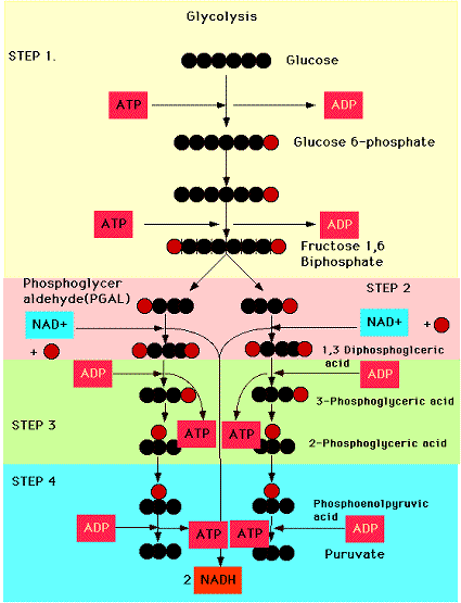
Anaerobic metabolism utilizes anaerobic respiration. This is performed through glycolysis, the process that converts glucose into pyruvate resulting in the creation of ATP. In this form of respiration, oxygen is not required. However, the utilization of anaerobic respiration for too long a time causes lactic acid build up, which results in the body becoming fatigued. Anaerobic metabolism is typically utilized by sprinters. Sprinters also take advantage of CP metabolism (also known as the ATP-CP metabolism or ATP-CP system), a process that first breaks down the ATP reserves in the body and then breaks down creatine phosphate to resynthesize ATP. Once the creatine phosphate runs out, then the sprinter returns to aerobic or anaerobic metabolism. The CP energy pathway allows for short bursts of intense energy for rapid movements.
Energy Pathway
Glucose to ATP, ADP and CP

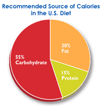
An athlete's diet is an essential part of their success. Since ATP and CP stores in the body are relatively small, glycogen, fat and protein stores allow for quick replenishment. Exercise physiologists suggest a diet of 55% carbohydrates which supply the glycogen stores which are used for short-term energy storage, 30% fats which are used for long-term energy storage, and 15% proteins, which, although they are not typically used as energy stores, they should be consumed to maintain the health of the muscles.

Suggested Diet Graph


Friday, October 29, 2010

Heart Surgery

The history of heart surgery is rather complex. The first surgery performed literally on the heart took place in Oslo in September 1895 when a surgeon ligated the coronary artery of a patient who was stabbed. Unfortunately, the patient passed away three days later. The first successful surgery on the heart was in September 1896 on a stabbed right ventricle. In 1925, Henri Souttar performed the first successful surgical treatment for mitral valve stenosis by palpating the damaged valve. It wasn't until after World War II that this method was adopted by surgeons across the United States. The first open heart surgery was performed in September 1952 by stopping and draining the blood of the heart.

Physical symptoms of an unhealthy heart can range from sweating to intense chest pains. The physiological signs of an underlying condition include an enlarged heart, more forceful heart beats and an abnormal heart rhythm. In order to detect a heart rhythm, doctors employ the use of an electrocardiogram (EKG or ECG). Since every time the heart beats electrical signals are set off by the nodes in the heart, these signals are recorded by the electrocardiogram which utilizes several wires with electrodes attached to the body to conduct these signals and record them on the monitor. A heartbeat is defined as every time the heart contracts and relaxes. The heart typically beats 70-80 times per minute at rest. However, due to the fact that a many patients reveal no abnormalities in a typical, resting electrocardiogram, a stress test (for example, having the patient run on a treadmill) is performed with the EKG still attached to record the heart when it is performing strenuous activity. There are five major points on an EKG revealed by each heartbeat, and they are labeled the P wave, QRS complex and T wave. When blood first entered the heart, it floods into the either the right atrium  (if it is oxygen-poor) or into the left atrium (if it is oxygen-rich). The sinoatrial node fires of a signal for the atria to contract, forcing the blood up into ventricles. This the P wave on the EKG. The QRS complex appears on an EKG when the atrioventricular node fires, causing the ventricles to contract. This contraction is called systole. The blood from the right ventricle is forced into the pulmonary artery and then the lungs to become oxygenated, while the blood from the left ventricle is forced through the aorta to flow to the rest of the body. When the ventricles contract, the tricuspid valve, the barrier between the right atrium and ventricle, and the mitral valve, the barrier between the left atriuim and ventricle, close so that the contraction of the ventricles does not force blood back the way it came, making a "lub" sound. The relaxation of the ventricles, called diastole, is exhibited by the T wave on the EKG. When the ventricles relax, the pulmonary and aortic valves close to prevent blood in these two major arteries from flowing backwards, making a "dub" sound.

EKG set-up
Placement of EKG electrodes

Blood flow through the heart

The heart and an EKG

There are three primary heart surgeries performed by cardiac surgeons: coronary bypass, heart transplant and angiocardiography. A coronary bypass the replacement of a damaged artery by removing a portion of the saphenous vein from the leg and placed where the diseased artery once was. A fun way to learn (and practice!) coronary bypass surgery is in this game courtesy of the Australian Broadcasting Corporation. Another excellent learning tool is created by a company called PlayGen which creates realistic simulations for professionals. A heart transplant is another major surgery performed by cardiac surgeons, during which the patient's damaged heart is removed and an artificial heart or a heart from a donor is placed in the chest cavity, and each of the arteries, veins and vessels are reattached. Angiocardiography is another procedure performed by cardiac surgeons. First, the doctor inserts a catheter into the arm or leg of the patient. He or she threads the catheter into the coronary artery and injects a dye which will fluoresce in an x-ray. This diagnostic tool allows the doctor to see if there are blockages in the artery, for if the dye flows through without a problem, the veins will appear to light up in the x-ray, but if there are such blockages, certain parts of each artery will appear dark for there is no flow of the dye through the veins.

One famous heart patient case history is that of Eileen Saxon more commonly known as "The Blue Baby." Saxon was born with the condition Tetralogy of Fallot, a severe congenital defect that prevents the blood from flowing through the heart properly, therefore resulting in the chronic lack of oxygen in the blood. This deoxygenated blood gave Saxon a blue appearance: her lips and fingers were blue and her skin had a bluish tinge. On November 29, 1944, which Saxon was only 15 months old, she underwent the first successful corrective surgery for the Tetralogy of Fallot by pioneering the Blalock-Taussig shunt which helped reroute the blood to be oxygenated. However, only a few months later did Saxon begin exhibiting symptoms of her congenital defect, and underwent another surgery, only to die a few days later, just before her third birthday. Although the surgery was not entirely successful, after the first shunt was placed, Saxon became a happy, pink, baby within two weeks of the surgery. The third time the surgery was peformed, this time was on a six-year-old boy, he began to turn pink quite quickly, establishing the Blalock-Taussing shunt as the premier corrective surgery for the Tetralogy of Fallot. For more information, visit the website from the hospital where it all took place, Johns Hopkins University, all due to Dr. Alfred Blalock, Dr. Heather Taussing, and assistant Vivien Thomas.


Tuesday, October 19, 2010

Artificial Organs and Regenerative Medicine

Recent discoveries made by biomedical engineers, and other scientists in related areas of research, have led to a new field of medicine, and new opportunities for patients with damaged organs. This field, regenerative medicine, is based off of the concept that every tissue in the human body has the capacity to regenerate, once cells that trigger cell division send the cues to the body. Currently, scientists are utilizing extracellular matrix, a combination of proteins and connective tissue, harvested from pig bladders and manipulated into powder form. This seemingly miraculous powder works on the cellular level to activate cells that build new cells when tissues become damaged. The field of regenerative medicine has proven quite promising; scientists have been able to trigger cell growth of 22 different types of tissues at one lab at Wake Forest University alone (for a comprehensive list of all the tissues, visit the Wake Forest Institute for Regenerative Medicine website). For example, these researchers were able to construct a completely functional esophagus and bladder. What was incredibly exciting was that, since cardiac cells are the only muscle cells that can move individually, the researchers at Wake Forest were able to build a heart valve that was already beating.

A researcher at Wake Forest University uses a mold to help an artificial bladder retain its shape and continue growing correctly


The benefits of artificial organs are obvious: fewer surgeries necessary, perhaps even a reduced need for organ transplants, lower risk of rejection because the organs are made from the body's own cells, the list of advantages are numerous. However, like any new scientific discovery, it has its pitfalls. First and foremost, since this breakthrough is so recent, there is not enough research that can conclude whether or not artificial organs can cause disorders or defects. Furthermore, the ultimate success of these organs are unknown, and treating a failing organ could be difficult. Another stumbling block is, similar to the debate of whether or not stem cell research is ethical, people are concerned by the ongoing research in these labs. Those in negation of this form of research question whether or not humans have the right to "play God" and create such organs.

Monday, October 4, 2010

Stem Cells

Currently, a major focus of biomedical researchers has been the study of stem cells. Stem cells can be categorized in three major groups. The first major group is known as embryonic stem cells (ES). These stems cells are derived, as their name suggests, from embryos that were formed in vitro. These cells can be further classified into totipotent and pluripotentThe totipotent stem cells are obtained from a fertilized egg in its earliest stages, whereas pluripotent stem cells are obtained from the blastocyst. These embryonic stem cells have not yet differentiated, thus, they can be used in any part of the body and become specific to a given tissue, organ, etc. This research as been hindered by the moral dilemma regarding whether or not a fertilized embryo is considered alive.

Totipotent and pluripotent stem cells
Another major category of stem cells is induced pluripotent stem cells (iPS). These cells are adult stem cells which are essentially "reprogrammed" to exhibit behaviors of an embryonic stem cell. They behave has pluripotent stem cells and can develop all of the germ layers of a normal cell, and then differentiate.

The final major category of stem cells is adult stem cells, also known as somatic stem cells. These cells are undifferentiated and are retrieved from mature tissues, as opposed to embryonic stem cells. While they can differentiate to be any type of cell, they typically differentiate to be the same type of cell as its surrounding tissue.
Cell differentiation
These cells can be manipulated to specialize in a laboratory. After retrieving a sample of the desired stem cells, the scientists then remove the outermost layer of the blastocyst, and allow them to foster in a petri dish. Colonies of the cells begin to grow, and as they do, certain cells begin to develop into endoderm, mesoderm and ectoderm cells. Scientists can then manipulate the differentiation by adding growth factors, causing the cell to believe that its environment is that of a specific tissue type. This process results in the differentiation of a cell.
Stem cells are invaluable to curing and treating various diseases. Currently, stem cells are being utilized to treat blood-cell disorders and in bone marrow. Ongoing research is studying the possibilities of using stem cells to treat damaged cardiac muscle in heart disease, creating skin grafts for burn victims, repairing the spinal cord, treating Parkinson's disease, Alzheimer's disease, diabetes, osteoarthritis, and rheumatoid arthritis.

Potential usage of stem cells to treat heart disease