The history of heart surgery is rather complex. The first surgery performed literally on the heart took place in Oslo in September 1895 when a surgeon ligated the coronary artery of a patient who was stabbed. Unfortunately, the patient passed away three days later. The first successful surgery on the heart was in September 1896 on a stabbed right ventricle. In 1925, Henri Souttar performed the first successful surgical treatment for mitral valve stenosis by palpating the damaged valve. It wasn't until after World War II that this method was adopted by surgeons across the United States. The first open heart surgery was performed in September 1952 by stopping and draining the blood of the heart.
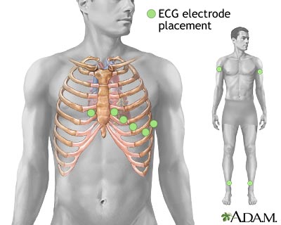
Physical symptoms of an unhealthy heart can range from sweating to intense chest pains. The physiological signs of an underlying condition include an enlarged heart, more forceful heart beats and an abnormal heart rhythm. In order to detect a heart rhythm, doctors employ the use of an electrocardiogram (EKG or ECG). Since every time the heart beats electrical signals are set off by the nodes in the heart, these signals are recorded by the electrocardiogram which utilizes several wires with electrodes attached to the body to conduct these signals and record them on the monitor. A heartbeat is defined as every time the heart contracts and relaxes. The heart typically beats 70-80 times per minute at rest. However, due to the fact that a many patients reveal no abnormalities in a typical, resting electrocardiogram, a stress test (for example, having the patient run on a treadmill) is performed with the EKG still attached to record the heart when it is performing strenuous activity. There are five major points on an EKG revealed by each heartbeat, and they are labeled the P wave, QRS complex and T wave. When blood first entered the heart, it floods into the either the right atrium (if it is oxygen-poor) or into the left atrium (if it is oxygen-rich). The sinoatrial node fires of a signal for the atria to contract, forcing the blood up into ventricles. This the P wave on the EKG. The QRS complex appears on an EKG when the atrioventricular node fires, causing the ventricles to contract. This contraction is called systole. The blood from the right ventricle is forced into the pulmonary artery and then the lungs to become oxygenated, while the blood from the left ventricle is forced through the aorta to flow to the rest of the body. When the ventricles contract, the tricuspid valve, the barrier between the right atrium and ventricle, and the mitral valve, the barrier between the left atriuim and ventricle, close so that the contraction of the ventricles does not force blood back the way it came, making a "lub" sound. The relaxation of the ventricles, called diastole, is exhibited by the T wave on the EKG. When the ventricles relax, the pulmonary and aortic valves close to prevent blood in these two major arteries from flowing backwards, making a "dub" sound.
There are three primary heart surgeries performed by cardiac surgeons: coronary bypass, heart transplant and angiocardiography. A coronary bypass the replacement of a damaged artery by removing a portion of the saphenous vein from the leg and placed where the diseased artery once was. A fun way to learn (and practice!) coronary bypass surgery is in this game courtesy of the Australian Broadcasting Corporation. Another excellent learning tool is created by a company called PlayGen which creates realistic simulations for professionals. A heart transplant is another major surgery performed by cardiac surgeons, during which the patient's damaged heart is removed and an artificial heart or a heart from a donor is placed in the chest cavity, and each of the arteries, veins and vessels are reattached. Angiocardiography is another procedure performed by cardiac surgeons. First, the doctor inserts a catheter into the arm or leg of the patient. He or she threads the catheter into the coronary artery and injects a dye which will fluoresce in an x-ray. This diagnostic tool allows the doctor to see if there are blockages in the artery, for if the dye flows through without a problem, the veins will appear to light up in the x-ray, but if there are such blockages, certain parts of each artery will appear dark for there is no flow of the dye through the veins.
One famous heart patient case history is that of Eileen Saxon more commonly known as "The Blue Baby." Saxon was born with the condition Tetralogy of Fallot, a severe congenital defect that prevents the blood from flowing through the heart properly, therefore resulting in the chronic lack of oxygen in the blood. This deoxygenated blood gave Saxon a blue appearance: her lips and fingers were blue and her skin had a bluish tinge. On November 29, 1944, which Saxon was only 15 months old, she underwent the first successful corrective surgery for the Tetralogy of Fallot by pioneering the Blalock-Taussig shunt which helped reroute the blood to be oxygenated. However, only a few months later did Saxon begin exhibiting symptoms of her congenital defect, and underwent another surgery, only to die a few days later, just before her third birthday. Although the surgery was not entirely successful, after the first shunt was placed, Saxon became a happy, pink, baby within two weeks of the surgery. The third time the surgery was peformed, this time was on a six-year-old boy, he began to turn pink quite quickly, establishing the Blalock-Taussing shunt as the premier corrective surgery for the Tetralogy of Fallot. For more information, visit the website from the hospital where it all took place, Johns Hopkins University, all due to Dr. Alfred Blalock, Dr. Heather Taussing, and assistant Vivien Thomas.


Nice to read your article! I am looking forward to sharing your adventures and experiences. famous cardiology hospital in Secunderabad
ReplyDeletehttps://getfitmississippi.blogspot.com/2012/05/cholestrol-lowering-tips-from-health.html?showComment=1641547138319#c4922949947352444998
ReplyDeleteHumulin insulin is a life-altering medication designed to assist individuals living with diabetes. Acting as an identical form of human insulin, it effectively reduces blood sugar levels in those affected by the disease. Humulin comes in four distinct forms: rapid-acting, short-acting, intermediate-acting and long-acting variations - all allowing patient's easy access to this life changing treatment through either injection or pump format!
ReplyDelete